⭐⭐⭐ UPPER GASTROINTESTINAL TRACT BLEEDING AND HAEMATEMESIS ⭐⭐⭐
⭐⭐⭐ UPPER GASTROINTESTINAL TRACT BLEEDING AND HAEMATEMESIS ⭐⭐⭐

1) DEFINITION OF HAEMATEMESIS :-
Hematemesis is the vomiting of blood. It is the bleeding proximal to duodenojejunal junction i.e ligament of treitz.

2) TREATMENT OF HAEMETEMESIS :- TREATMENT IS IMPORTANT FIRST .
(I) Primary treatment -
# Patient's condition should be made stable first. Monitor Pulse, BP , respiration, consciousness, Urine output. Blood sample is taken for Hb testing.
# Body fluid replenishment by fluids & blood. ( Hb should be kept > 7-8 gm/dl.)
(II) Gastric lavage -
# Nasogastric tube (Ryle's tube) is inserted & 500 ml tap water is instilled & aspirated every 30-60 mins.
# Vasoconstriction occurs due to cool water which helps in temporary cessation of bleeding.
# It helps to clear stomach prior to endoscopy, assess the amount of bleeding.
# Forceful suction can damage mucosa.
# Tube is kept for 24 hours to assess chances of rebleeding.
(III) Endoscopy :-
# It helps to diagnose the cause, assess chances of rebleeding.
# Massive bleeding in varices is diagnosed by Large varices, Daughter varices, Cherry Red Spots on varices.
# Massive bleeding in peptic ulcer is diagnosed by Sentinel clot on surface of ulcer, exposed vessels in the base of ulcer.
(IV) Other investigations:-
# Angiography & radionuclide scanning-
To detect bleeding site.
(V) Other treatment :-
# PPI (proton pump inhibitor) - Omeprazole 40 - 80 mg i.v. followed by 8 mg per hourly.
PPIs reduce gastric acid secretion by inhibiting H+, K+ ATPase pump.
This promotes healing of ulcers and erosions as well as stabilizing thrombi and decreasing rates of GI bleeding .
(A gastric acidic environment alters coagulation function and activates pepsin to break the platelet plugs. Hence, PPI decrease bleeding by reducing acid and promoting thrombosis)
# FFP & platelet transfusion - for cases with active bleeding & thrombocytopenia.
# Antifibrinolytic agent - Tranexamic acid. - it prevents the lysis of thrombus and hence decreases bleeding.
# Balloon tamponade, Vasopressin. ( cause vasoconstriction)
- for varices.
# Therapeautic endoscopy -
Thermal t/t- Laser, electrocoaguation.
Non thermal t/t - Endoscopic sclerotherapy, band ligation of bleeding vessels.
# Radiotherapy.
# Surgery - Done when medical measures fail.
For Varices - Shunt surgery or transaction (devascularization of varices).
For Gastric erosions - Gastrectomy, vagotomy with drainage. (Since vagus stimulates parasympathetic system of stomach & thereby increase gastric secretion, vagotomy will prevent the secretion & further gastric erosion).
🌺🌺🌺🌺🌺
3) INVESTIGATIONS OF HAMETEMESIS :-
(I) Endoscopy. helps to diagnose the cause, assess chances of rebleeding.
(II) Angiography. Helps to detect site of bleeding
(III) Nasogastric aspiration.
(IV) ECG - if possibility of heart diseases.
(V) Blood tests -
# Hb.
# Hematocrit.
# Coagulation Profile.
# LFT.
(Liver produces coagulation factors, liver disease can also lead to portal Hypertension and oesophageal varices which may bleed.)
# Platelet Count.
# Urea & Creatinine.
Uremia due to chronic kidney disease may cause haematemesis. Explained later in this article
🌺🌺🌺🌺🌺
4) COMPLICATIONS OF HAEMATEMESIS:-
(I) Shock - hypovolemic shock.
Symptoms of shock :-
- cold or clammy skin
- pale skin
- rapid, shallow breathing
- rapid heart rate
- little or no urine output
- confusion
- weakness
- weak pulse
- blue lips and fingernails
- fainting and loss of consciousness
(II) Pallor
(III) Angina - due to decreased blood supply to the heart.
(IV) Syncope - due to decreased blood supply to the brain.
(V) Hypotension, tachycardia
🌺🌺🌺🌺🌺
5) HOW TO FIND OUT POSSIBLE CAUSE OF HAEMATEMESIS :-

(A) OESOPHAGEAL CARCINOMA :-
- At first, a cancer may bleed slightly because its blood vessels are fragile. Later, as the cancer enlarges and invades surrounding tissues, it may grow into a nearby blood vessel, causing bleeding.
- Note :- Below points are ONLY FOR INFORMATION
Oesophageal carcinoma can be suspected if any of the following features are present:-
# Dysphagia
(when the lumen is filled with cancer tissue)
# Anorexia and weight loss -
(In an effort to fight the cancer, the body produces different cytokines. These cytokines can lead to weight loss, muscle loss, and a decrease in appetite.)
# Substernal and abdominal pain -
(Cancer tissue invades and damages nearby tissues and nerves causing pain. It also leads to release of some mediators and cytokines which cause pain.)
# Palpable supraclavicular lymph nodes-
(due to spread of carcinoma to the lymph node.)
# Back pain
( if the cancer invades pericardium or mediastinum.)
# Hoarseness
(recurrent laryngeal nerve involvement) or hiccups ( phrenic nerve involvement)
# Liver secondaries -
( As the secondaries in the liver advance, the liver may swell.)
# Ascites :-
(Spread of cancer to liver causes blocking of portal venules resulting in increased pressure and fluid leakage.
Cancer can also spread to the peritoneum and make it leaky, causing malignant ascites.)
# Bronchopneumonia -
(It can occur if food enters the lungs through trachea because a tumor is blocking the esophagus .)
Investigations of oesophageal carcinoma:-
- Barium swallow
- Endoscopy
- Biopsy
- CT, PET scan ( for extent of tumor)
⭐⭐⭐⭐
(B) OESOPHAGITIS
- Inflammation causes damage to the small vessels and bleeding.
Note :- Below points are ONLY FOR INFORMATION
- Oesophagitis can be suspected if any of the following features are present:-
# Painful swallowing
( due to damaged nerve endings and inflammatory mediators.)
# Heartburn
(due to Inflammation)
# Acid regurgitation
(bringing food back up to the mouth from the stomach due to damage to the lower esophageal sphincter which usually keeps the acidic contents of the stomach out of the esophagus. ) continuous cycle of acid reflux causes scarring and narrowing of the esophagus.
- Causes of oesophagitis :-
# Backflow of acid from the stomach to the esophagus (GERD)
Following Investigations may help to diagnose:-
Oesophageal manometry- checks function
of oesophageal sphincter.
24 hour pH monitoring
Endoscopy, mucosal biopsy
Barium study.
# Vomiting -
Ask about history of Vomiting and illness.
when vomiting is frequent or chronic it can lead to acid damage to the esophagus.
# Medications such as aspirin and NSAIDS - Ask about history of these medications.
it causes esophagitis by decreasing prostaglandins. Prostaglandins (PGs) protect the esophagus by inhibiting acid secretion, stimulate mucus and bicarbonate secretion, alter mucosal blood flow.
# Viruses, fungi, bacteria, or diseases that weaken the immune system e.g HIV
- Look for fever, history of any medications, perform tests like CBC.
⭐⭐⭐⭐
(C) MALLORY WEISS SYNDROME (MWS) :-
- It is the tear or laceration of the mucous membrane, most commonly at the point where the esophagus and the stomach meet (gastroesophageal junction).
Such a tear may result in severe bleeding.
Note :- Below points are ONLY FOR INFORMATION
- MWS can be suspected if any of the following features are present:-
# Abdominal pain -
(due to tearing of nerves and also the blood released is very irritating to the tissues. )
# vomiting up blood i.e hematemesis
due to bleeding from the torn vessels.
# involuntary retching
( making the sound and movement of vomiting . The diaphragm contracts when trying to vomit but the body is unsuccessful in doing so.)
# bloody or black stools ( melena)
(blood from tear goes into Intestine)
- Investigation of MWS :-
# Endoscopy
# Stool examination (blood in stool)
- Causes of MWS :-
# Severe , forceful and prolonged vomiting causes trauma and tear.
Ask about history of Vomiting
Stomach illness
Chronic alcohol abuse - ask about history
# Trauma to the chest or abdomen
Ask about history of trauma.
# Severe or prolonged hiccups -
Ask for history of hiccups.
(Hiccups are periodic contractions of the diaphragm and it could exert shear forces on the gastroesophageal junction and tear the mucosa.)
# Intense coughing can cause trauma and tear. Ask for history of cough.
# heavy lifting or straining .Ask for history.
It can cause pressure and tear.
# gastritis -
(inflammation can cause damage and tear.)
Look for additional symptoms like belching, heartburn, indigestion, loss of appetite.
Tests :- tests for H. pylori , endoscopy.
# Hiatal hernia,
(It occurs when part of your stomach pushes through part of your diaphragm - tear can occur because of trauma to the herniated part at the diaphragmatic opening or due to acid reflux.)
Tests include:-
Barium swallow test, endoscopy , esophageal manometric studies, pH test , gastric emptying studies.
# convulsions:- Ask about history of convulsions.
# Receiving cardiopulmonary resuscitation (CPR) can also lead to a tear due to trauma.
ENDOSCOPY AND STOOL EXAMINATION (MELENA) MAY HELP IN DIAGNOSIS OF MALLORY WEISS SYNDROME.
⭐⭐⭐⭐
(d) OESOPHAGEAL VARICES OR COLLATERALS :- OCCURS IN PORTAL HYPERTENSION.
- Esophageal varices develop when normal blood flow to the liver from portal venous system is blocked by a clot or scar tissue in the liver leading to portal hypertension.
To bypass the blockages, blood flows into smaller blood vessels that aren't designed to carry large volumes of blood.
The vessels can leak blood or even rupture, causing life-threatening bleeding.
Note :- Below points are ONLY FOR INFORMATION
- esophageal varices can be suspected if any of the following features are present :-
( Some of these may be present while
some may not be present)
# Hematemesis and Melena
(due to bleeding from varices.)
# Caput medusae-
( collaterals around umbilicus. )
# Ascites, anasarca, hydrothorax -
(due to portal hypertension, hydrostatic pressure in veins is increased causing leakage of fluid outside.)
# Splenomegaly -
Congestive splenomegaly due to backpressure of blood in splenic vein (which is a tributary of portal vein) due to blockage of blood flow from portal system to liver .
# Venous hum -
(sound produced due to turbulent blood flow in veins due to increased pressure - heard more during inspiration in epigastric region.)
# Hemorrhoids ( piles) -
due to rectal varices.
# Foetor hepaticus -
musty odour of breath - this is due to the mercaptans reaching the lungs directly as liver is bypassed due to collaterals.
# General features -
- Jaundice - in case of hepatic
damage.
- Pruritus - In case of hepatic cause
( Liver disease), cholestasis causes
rupture of bile ductules and entry
of bile salts in blood which further
accumulate under skin causing
itching.
- Abdominal pain -
due to swollen liver or nerve irritation because of ascites or intraabdominal bleeding from varices ,etc.
- Anorexia , weight loss - due to
decreased food intake or improper
digestion of food when bile is
decreased.
- fatigue
- Hypotension -
( Splanchnic circulation- arterial
branches which supply git, liver,
spleen, pancreas . These arterial
branches further form capillaries
and terminate in portal venules. )
Portal hypertension causes
splanchnic vasodilation due to
release of vasodilators like Nitric
oxide. Splanchnic vasodilation
causes hypotension.
- Cyanosis - due to portopulmonary
anastomosis with flow of blood in
direction of pulmonary veins.
- Muscle cramps - common in
cirrhosis
- Bleeding, bruises - inadequate
clotting factor production in case
of liver damage.
# Signs of liver failure -
(Refer to the article of portal hypertension for explanation)
- Gynaecomastia ,
- Testicular atrophy,
- Palmar erythema ,
- spider nevi,
- parotid enlargement,
- hepatorenal syndrome, - hepatopulmonary syndrome,
- cardiomyopathy, osteodystrophy,
hepatic encephalopathy , etc
# Liver consistency - if firm - cirrhosis
# Spontaneous bacterial peritonitis-Due to ascitic fluid infection. There is pain, fever, raised TLC, positive ascitic fluid culture.
# Previous history of risk factors like
Alcohol, OC pills ,etc
- IF ANY OF THE ABOVE SYMPTOMS ARE SEEN WITH HAEMATEMESIS, FOLLOWING INVESTIGATIONS MAY BE CARRIED OUT TO DIAGNOSE VARICES:-
(A) BARIUM SWALLOW :- varices in
Oesophagus - bag of worm
appearance
✨✨✨
(B) UPPER G.I SCOPY - to see oesophageal varices.
✨✨✨
(C) LFT i.e LIVER FUNCTION TEST
(I) TB
(increase in ALP, serum globulin but normal transaminase )
TB causes inflammation and fibrosis of portal venules, sinusoids which obstructs the flow of blood and causes portal hypertension and varices.
There may be mild fever, right upper quadrant pain, hepatomegaly, weakness, night sweats, weight loss, jaundice, liver tenderness.
Look for previous medical history of tuberculosis or contact with tuberculosis pt. Elevated levels of ESR, positive tuberculin test;
🌟🌟
-(II) Sarcoidosis -
Increase in liver enzymes alkaline phosphatase and gamma glutamyl transpeptidase
In Sarcoidosis, Inflammation and granuloma formation occurs which obstructs the flow of blood and causes portal hypertension and varices.
Itching may be present.
🌟🌟
-(III) Liver cirrhosis
Signs of liver cell failure may be seen. ( Refer to the article of portal hypertension for explanation of signs )
🌟🌟
-(IV) Fatty liver
(too much fat build up in your liver)
Following symptoms may be present:- Abdominal pain or a feeling of fullness in the upper right side of the abdomen, Swollen abdomen, edema in legs ( due to decreased protein production by liver)
Nausea, loss of appetite, jaundice, fatigue.
✨✨✨
(D) USG - (ULTRASOUND)
- ascites,
- liver or spleen enlargement,
- varices
- Pressure on portal vein due to stomach or pancreas - leading to obstruction to blood flow in portal vein and causing portal hypertension.
✨✨✨
(E) PORTAL VENOGRAPHY-
Portal venous thrombosis or splenic venous thrombosis
✨✨✨
(F) IMAGING METHODS LIKE
CT, MRI , ANGIOGRAPHY, DOPPLER USG
(I) Portal venous thrombosis or splenic venous thrombosis
It causes portal hypertension which further results in varices and haematemesis.
(II) Periportal inflammation
Inflammation leads to fibrosis and
narrowing of lumen of vein which causes portal hypertension .
(III) Congenital hepatic fibrosis
Congenital malformation of portal venules causes obstruction of portal venules and portal hypertension.
(IV) TB
Inflammation and fibrosis of portal venules, sinusoids.
There may be mild fever, right upper quadrant pain, hepatomegaly, weakness, night sweats, weight loss, jaundice, liver tenderness.
Look for previous medical history of tuberculosis or contact with tuberculosis pt. There are elevated levels of ESR, positive tuberculin test;
(V) Sarcoidosis - inflammation and granuloma formation occurs which obstructs the flow of blood and causes portal hypertension and varices.
Itching may be present.
(VI) Wilson's disease
Copper builds up in liver - damage and portal hypertension.
Additional symptoms may be:-
- Golden-brown eye discoloration (Kayser-Fleischer rings)
- Problems with speech, swallowing or physical coordination.
- Uncontrolled movements or muscle stiffness.
(VII) Liver cirrhosis
Signs of liver cell failure may be seen. ( Refer to the article of portal hypertension for explanation of signs )
(VIII) Hepatitis
Alcoholic or viral hepatitis leads to inflammation of liver which damages the liver sinusoids and causes portal hypertension.
Following symptoms may be present:-
Swollen liver, jaundice, fever in prodromal phase, etc.
(IX) Fatty liver
(too much fat build up in your liver which damages the liver sinusoids and causes portal hypertension.)
Following symptoms may be present:-. Abdominal pain or a feeling of fullness in the upper right side of the abdomen, Swollen abdomen, edema in legs ( due to decreased protein production by liver)
Nausea, loss of appetite, jaundice, fatigue.
(X) Polycystic liver disease. It damages the liver sinusoids and causes portal hypertension.
Ask for family history.
There may be swelling in the abdomen when liver enlarges.
(XI) Pulmonary hypertension
Pulmonary hypertension causes increased backpressure in right heart and then into IVC . Backpressure in IVC leads to increased pressure in hepatic vein and then in portal vein causing portal hypertension.
Symptoms may be:-
- Shortness of breath
(Pulmonary hypertension can lead to accumulation of blood in the lung which leads to development of pleural effusions and pulmonary edema)
- Fatigue (decreased cardiac output)
- Dizziness or fainting (due to decreased amount of blood reaching left heart and thus decreased cardiac output)
- Chest pressure or pain
- Swelling (edema) in the ankles, legs and eventually the abdomen (ascites) due to backpressure in veins.
- cyanosis
- tachycardia (decreased cardiac output)
(XII) Hemochromatosis
Excessive iron is stored in liver causing damage.
Symptoms may be:-
Joint pain, Abdominal pain ,Fatigue,
Impotence, Heart failure, Liver failure
Bronze or gray skin color ,Memory loss.
(XIII) Congestive heart failure (CT, MRI)
Heart failure leads to backpressure in IVC due to inability to pump. Backpressure in IVC leads to increased pressure in hepatic vein and then in portal vein causing portal hypertension.
There may be symptoms like
- Shortness of breath (due to decreased blood supply to lungs)
- Fatigue (due to decreased blood supply to body)
- Swelling in the legs, ankles and feet due to backpressure in veins.
- Rapid or irregular heartbeat
- Persistent cough or wheezing with white or pink blood-tinged mucus (due to increased pressure in pulmonary veins)
- Swelling of the abdomen
- Nausea
- Chest pain
(XIV) Inferior Vena Cava thrombosis
Backpressure in IVC leads to increased pressure in hepatic vein and then in portal vein causing portal hypertension.
Symptoms may be as follows:-
- hypotension ( due to decreased venous return and hence decreased cardiac output)
- tachycardia ( decreased cardiac output causes hypoxia in tissues, hence heart compensates by beating rapidly)
- lower extremity edema, ascites (due to increased pressure in veins)
elevated liver enzymes,
end-organ failures and, hypoxia, altered mental statusstatus
- pallor, shortness of breath , fatigue, cold extremities (decreased cardiac output)
- diaphoresis, dizziness, altered mental status ( Decreased blood flow to brain)
(XV) Budd Chiari syndrome - Hepatic vein thrombosis.
( It causes portal hypertension which further results in varices and haematemesis.)
The symptoms of Budd-Chiari syndrome include:
- Ascites (swelling in the abdomen caused by increased hydrostatic pressure in vein.)
- Jaundice
- Enlarged and tender, painful liver.
- Edema (swelling) in the legs.
- Hepatic encephalopathy
- Vomiting.
- Enlarged spleen.(congestive Splenomegaly )
✨✨✨✨
(G) LIVER BIOPSY
- Sarcoidosis,
inflammation and granuloma formation occurs which obstructs the flow of blood and causes portal hypertension and varices.
Itching may be present.
- Wilson disease,
Copper builds up in liver - damage and portal hypertension.
Additional symptoms may be:-
- Golden-brown eye discoloration (Kayser-Fleischer rings)
- Problems with speech, swallowing or physical coordination.
- Uncontrolled movements or muscle stiffness.
- hepatitis,
Alcoholic or viral hepatitis leads to inflammation of liver which damages the liver sinusoids and causes portal hypertension.
Following symptoms may be present:-
Swollen liver, jaundice, fever in prodromal phase, etc.
- Hemochromatosis
Excessive iron is stored in liver causing damage.
Symptoms may be:-
Joint pain, Abdominal pain ,Fatigue,
Impotence, Heart failure, Liver failure
Bronze or gray skin color ,Memory loss.
✨✨✨✨
(H) ASCITIC FLUID STUDY .
✨✨✨✨
(I) STOOL AND URINE MICROSCOPY
- Schistosomiasis
( Look for Fever, Abdominal pain , Bloody diarrhea ,Cough Rash, Body aches and headache, serum antibodies.)
✨✨✨✨
(J) BLOOD TEST :-
- Wilson's disease, (for explanation, see above)
- hepatitis, (for explanation, see above)
- Hemochromatosis, (for explanation, see above)
- Pulmonary hypertension (for explanation, see above)
✨✨✨✨
(K) ECG, ECHOCARDIOGRAM, CARDIAC CATHETERIZATION -
- Congestive heart failure, (for explanation, see above)
- right heart failure, (for explanation, see above)
- tricuspid insufficiency
- Pulmonary hypertension (for explanation, see above)
✨✨✨✨
(L) CHEST X RAY , PULMONARY FUNCTION TESTS :- Pulmonary hypertension
(for explanation, see above)
⭐⭐⭐⭐
(e) TRAUMA TO OESOPHAGUS :- can cause haematemesis. There is history of trauma.
Inv :- Esophagography with a water-soluble contrast agent, helps in diagnosis.
⭐⭐⭐⭐⭐⭐⭐⭐⭐⭐⭐⭐⭐⭐⭐
(II) GASTRODUODENAL CAUSES OF HAEMATEMESIS :-
(A) EROSIVE GASTRITIS OR DUODENITIS :-
It is characterized by many inflamed lesions in the mucous lining of the stomach. This inflammation damages blood vessels leading to haematemesis.
Note :- Below points are ONLY FOR INFORMATION
Other symptoms of gastritis/ duodenitis:-
- Pain in stomach,
- Nausea, vomiting, loss of appetite and weakness.
- In severe cases there can be haematemesis which may result in anemia.
- belching, heartburn, indigestion
Investigations include :-
- upper GI endoscopy
- History of alcohol,
( after repeated alcohol use begins to irritate or even erode parts of the stomach lining. This, in turn, can leave the stomach lining more vulnerable to the acidic digestive juices normally produced by the body to digest food.)
- History of stress and anxiety
(stress response results in atrophy of the gastric mucosa. In the stressed state, elevate levels of Acetylcholine and histamine result in increased acid production, thus inducing gastritis. Blood flow to the stomach also decreases )
- History of persistent use of aspirin or NSAID medications such as ibuprofen.
( NSAIDs cause damage to the gastroduodenal mucosa by causing irritation on the epithelium, impairment of the barrier properties of the mucosa, suppression of gastric prostaglandin synthesis, reduction of gastric mucosal blood flow )
- Stool antigen test and urea breath test , blood test - Infection like H pylori can cause gastritis.
- Intestinal endoscopy - Crohn’s disease (inflammatory bowel disease):-
Crohn's disease may present with following additional symptoms along with symptoms of gastritis:-
- Diarrhea
- Fever
- Blood in stool
- Mouth sores
- Reduced appetite and weight loss
- Anal fistula
⭐⭐⭐⭐
(B) STRESS ULCERS:-
are multiple, superficial mucosal erosions which occur mainly in the fundus and body of the stomach due conditions of stress like serious injury , infection , shock, sepsis, trauma , peritonitis and other chronic medical illness like heart failure, liver disease, kidney failure, stroke, hypertension, previous gastrointestinal disease and treatment with corticosteroids, NSAIDS.
This inflammation and erosion damages blood vessels leading to haematemesis.
Note :- Below points are ONLY FOR INFORMATION
Other symptoms of a stress ulcer include:
- pain in the upper stomach
- bloating
- nausea or vomiting (Peptic ulcers block passage of food through the digestive tract, either through swelling from inflammation or through scarring thus causing vomiting.)
- symptoms of anemia,
- Some ulcers bleed a lot, causing dangerous blood loss. In people who are already fighting serious injuries, this blood loss can be life-threatening.
- Symptoms of a rapidly bleeding ulcer include:
red or coffee grounds vomit
red or dark tarry stools
fainting ( due to hypotension)
Pathogenesis of stress ulcer - In conditions of stress like those given above, there is reduction in mucosal blood flow or a breakdown of normal mucosal defense mechanisms along with the injurious effects of acid and pepsin on the gastroduodenal mucosa.
History of chronic illness and situations of stress given above favours the diagnosis of stress ulcer. Endoscopy can also be done.
⭐⭐⭐⭐
(C) PEPTIC ULCER ( GASTRIC OR DUODENAL):-
lesion in the mucosa of the stomach or duodenum, caused by the action of pepsin and stomach acid. Erosion of blood vessels causes haematemesis.
Note :- Below points are ONLY FOR INFORMATION
Other symptoms of a peptic ulcer include:
- Pain in upper abdomen
- belching, heartburn, indigestion, passing excessive amounts of gas,. ( bacteria in your small intestine can lead to bloating from excess production of gas)
- vomiting
- fatigue, feeling full sooner than normal, or loss of appetite
Investigations of peptic ulcer:-
- Laboratory tests for H. pylori - blood, stool or breath test.
- Endoscopy :- biopsy may be removed for examination.
- barium swallow
- History of chronic intake of medications like aspirin, ibuprofen, and other NSAIDS.
- History of smoking or alcohol, radiotherapy.
- Stomach cancer (CT scan, barium swallow, upper endoscopy.)
⭐⭐⭐⭐
(D) GASTRIC CARCINOMA :-
Causes of bleeding in gastric carcinoma are tumors ulcers , varices, etc. Blood vessels of tumor are fragile and they bleed easily. Further, tumor invades the surrounding blood vessels causing bleeding.
Note :- Below points are ONLY FOR INFORMATION
Other symptoms of a gastric carcinoma include :-
- Difficulty swallowing
(when the lumen of stomach is filled with cancer tissue)
- Feeling bloated after eating
- Feeling full after eating small amounts of food
- Heartburn
(due to Inflammation in the tumor mass)
- Indigestion
- Vomiting
(particularly vomiting up solid food shortly after eating ·This occurs due to inability of the stomach to digest the food due to tumor invasion.)
- Stomach pain
(Cancer tissue invades and damages nearby tissues and nerves causing pain. It also leads to release of some mediators and cytokines which cause pain.)
- Unintentional weight loss and anorexia
(In an effort to fight the cancer, the body produces different cytokines. These cytokines can lead to weight loss, muscle loss, and a decrease in appetite. Fatigue is often related to anemia due to blood loss in the stool.)
Investigations of gastric carcinoma:-
- Barium swallow
- Endoscopy
- Biopsy
- CT, PET scan ( for extent of tumor)
⭐⭐⭐⭐
(E) HEMOBILIA :-
bleeding from and/or into the biliary tract
Causes:-
- Iatrogenic :- History of any invasive procedures involving the liver, pancreas, bile ducts or the hepatopancreatobiliary vasculature,
- trauma - History of trauma
- Other causes include
gallbladder and bile duct stones, biliary varices, biliary parasites (eg, Ascaris ), benign and malignant tumors involving the biliary tree, vascular aneurysms, pancreatitis, and hepatitis.
It is characterized by classic triad of right upper quadrant pain, jaundice, and overt upper gastrointestinal bleeding , hemobilia causes melena or haematemesis, acute biliary pain from distension of the bile ducts , jaundice
Investigations of Hemobilia:-
Magnetic resonance cholangiography or transabdominal ultrasound , CT scan.
⭐⭐⭐⭐
(F) INVASIVE PANCREATIC CARCINOMA:-
Pancreatic adenocarcinoma directly invading duodenum complicated by hemorrhage can be a rare cause of haematemesis.
Clinical features of pancreatic cancer:-
- Pain in the abdomen or middle back
- ascites
( due to obstruction of diaphragmatic lymphatics, increased production of exudate by the tumor itself and production of osmotically active peptides that alter vascular permeability to favor ascites formation )
- nausea, vomiting
cancer may block the duodenum due to which food can't pass down.
- fatigue , loss of appetite, weight loss
- dark urine, or yellow skin and eyes
(Pancreatic cancer that blocks the liver's bile duct can cause jaundice. )
Investigations of pancreatic cancer:-
- CT scans, MRI
- biopsy
- Tumor marker test used in pancreatic cancer is called CA19-9. It may be helpful in understanding how the cancer responds to treatment.
- Amylase and/or lipase—blood levels of these pancreatic enzymes may be elevated in people with pancreatic cancer.
⭐⭐⭐⭐⭐⭐⭐⭐⭐⭐⭐⭐⭐⭐⭐
(III) MISCELLANEOUS CAUSES OF HAEMATEMESIS :-
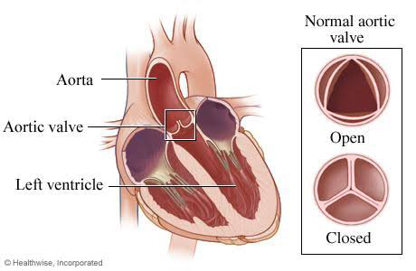
(A) AORTIC ANEURYSM :-
An aneurysm occurs when a section of the artery wall weakens and forms a bulge that can expand over time. An aneurysm can rupture if it experiences enough stress, causing fatal bleeding . Rupture of an abdominal aortic aneurysm into the duodenum through aortoduodenal fistula (ADF) leads to haematemesis.
Note :- Below points are ONLY FOR INFORMATION
Other symptoms of aortic aneurysm include :-
- Tenderness or pain in the chest. In case of abdominal aneurysm ,there is deep, constant pain in the abdomen or side of the abdomen or back pain .
Aorta receives sensory innervation from the aortic nerve. Dilatation of aorta causes stretching of the walls and stimulation of these nerves causing pain.
- Hoarseness
Direct compression of the enlarging thoracic aneurysm on the left recurrent laryngeal nerve causes injury to the nerve, which results in hoarseness.
- Cough
It is secondary to bronchial compression due to aneurysm.
- Shortness of breath
Thoracic aortic aneurysms may produce breathlessness by compressing the tracheobronchial tree.
Signs and symptoms of ruptured aneurysm include:
- Sudden, intense and persistent chest or back pain.
Nerves in the area of ruptured wall cause pain. Blood also causes irritation of nerves.
Also, the abdominal aorta is present deep in the abdomen and near the front of the spine. Because the aorta is near the spine, pain is felt in the back when aneurysm ruptures.
- Low blood pressure (due to loss of blood)
- Loss of consciousness (due to decrease in blood flow to brain)
- Shortness of breath
(due to loss of blood, there is decreased blood flow to lungs and brain 🧠 resulting in decreased oxygen supply to the tissues. This causes dyspnea.)
- Trouble swallowing
thoracic aorta aneurysm compresses to the esophagus, causing difficulty in swallowing.
- Weakness or paralysis of one side of the body, difficulty speaking, or other signs of a stroke ( due to decrease in blood flow to brain)
Investigations of ruptured aneurysm:-
First stabilize the vitals of patient
- rapid bedside ultrasonography (US) - Doppler study of abdominal aorta
- CT scanning, and MRI.
Causes of Aortic aneurysm:- (weakening of wall due to damage to the wall)
- Atherosclerosis
It occurs when fat and other substances build up on the lining of a blood vessel forming atherosclerotic plaque. This plaque can also burst, leading damage to arterial wall.
Investigations :- lipid profile
- High blood pressure.
High blood pressure can cause direct damage and weaken the aorta's walls.
Check BP
- Blood vessel diseases.
These are diseases that cause blood vessels to become inflamed. E.g autoimmune diseases, connective tissue disorder.
Investigation :-
Blood tests may show abnormal levels of certain blood cells and antibodies.
Biopsy , angiography.
C-Reactive Protein (CRP), ESR, ELISA
- Infection in the aorta.
Rarely, a bacterial or fungal infection might cause aneurysm by causing inflammation and damage to aortic wall.
Investigations :- CBC
- Trauma.
- Tobacco use and family history are risk factors.
⭐⭐⭐⭐
(B) COAGULATION DEFECTS:- e.g hemophilia
Along with haematemesis, it may present with other symptoms like melena, hematochezia, hemoptysis, easy bleeding from wound, etc
Investigations of coagulation defect:-
- bleeding time (BT) ,
- platelet count,
- activated partial thromboplastin time (aPTT),
- prothrombin time (PT), and thrombin time (TT).
- tests for clotting factors
- LFT (to check the function of liver. Liver produces clotting factors. In liver disease, production of clotting factors is decreased.)
⭐⭐⭐⭐
(C) SWALLOWED BLOOD IN CASE OF EPISTAXIS AND HEMOPTYSIS OR BLEEDING GUMS CAN LEAD TO HAEMATEMESIS.
Swallowed blood can irritate your stomach and cause vomiting.
⭐⭐⭐⭐
(D) CHRONIC INTAKE OF DRUGS LIKE ANTICOAGULANTS
( decreases clotting of blood)
NSAIDS
(NSAIDs increase bleeding by decreasing the activity of blood platelets and therefore decreasing formation of blood clots ) ,
STEROIDS
(corticosteroids may impair tissue repair, thus leading to delayed wound healing)
Ask about history of intake of these medications.
⭐⭐⭐⭐
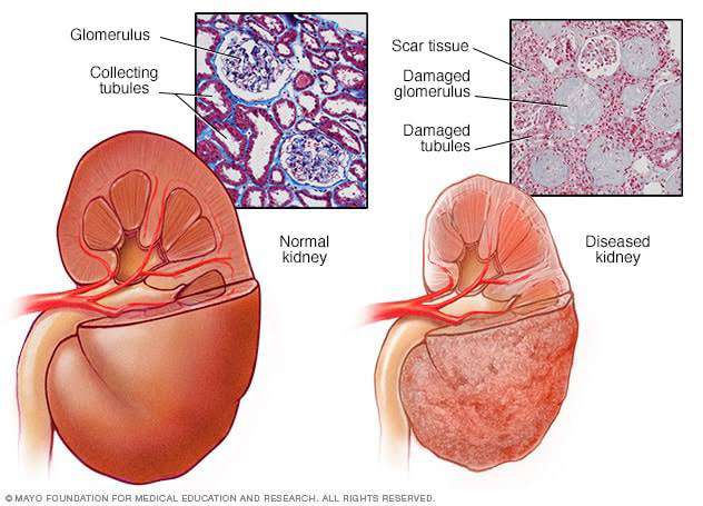
(E) UREMIA :-
There is raised level in the blood of urea and other nitrogenous waste compounds that are normally eliminated by the kidneys. Uremia is caused due to chronic kidney disease.
It causes platelet abnormalities. This leads to bleeding.
Note :- Below points are ONLY FOR INFORMATION
Other symptoms of Uremia:-
- extreme tiredness or fatigue, cramping in your legs
Uremic toxins induce apoptosis. Uraemic toxins impair skeletal muscle regeneration by inhibiting myoblast proliferation, reducing myogenic differentiation, and promoting muscular fibrosis
- little or no appetite
Accumulation of anorexigenic compounds, inflammatory cytokines, high brain serotonin levels and a lower serotonin/dopamine ratio cause anorexia.
( Urea is produced in the liver and is a breakdown product of amino acids.
Plasma and brain amino acids are involved in serotonin synthesis and appetite control. )
- headache
abnormal increase of non-protein nitrogen in the blood
- vomiting
accumulation of metabolic substances in the blood causes the vomiting
- Trouble concentrating, mental status changes
accumulation of Uremic toxins is responsible for vascular endothelial dysfunction.
exposure of both macrophages and astrocytes to Uremic toxins causes the release of inflammatory cytokines and oxidative stress in the brain microenvironment. These deleterious mechanisms, together with the direct neurotoxic properties of certain UTs, promote neuronal death.
- cardiac arrhythmias
due to electrolyte disturbances
Investigations of Uremia :-
- RFT (blood urea, serum creatinine, sodium, potassium, uric acid)
- Urinanalysis
⭐⭐⭐⭐
(F) HEREDITARY HEMORRHAGIC TELANGIECTASIA :-
Rare autosomal dominant genetic disorder that leads to abnormal blood vessel formation in the skin, mucous membranes, and often in organs such as the lungs, liver, and brain. This can cause bleeding.
Signs and symptoms of HHT include:
- Nosebleeds, sometimes on a daily basis and often starting in childhood
- red vessels or tiny red spots, particularly on the lips, face, fingertips, tongue and inside surfaces of the mouth
- Iron deficiency anemia
- Shortness of breath (due to anemia)
- Headaches
- Seizures (intracranial bleeding)
Investigations of Hereditary hemorrhagic telangiectasia :-
- Genetic testing
⭐⭐⭐⭐⭐⭐⭐⭐⭐⭐⭐⭐⭐⭐⭐
🌺🌺🌺🌺🌺

The content was really good and informative.. Looking forward to more blogs
ReplyDeleteThank you so much 😊😊
DeleteImpressive! Pointwise explanation of each n every heading is the speciality of your writing. Very good👌👌
ReplyDeleteThank you so much 😊😊
DeleteGreAt
ReplyDeleteThank you so much 😊😊
Delete👌👌👌
ReplyDeleteThank you so much 😊😊
DeleteVery good
ReplyDeleteThank you so much 😊😊
DeleteThank you so much 😊😊
ReplyDeleteBeta awesome👌👌
ReplyDeleteखुप दिवसांनी लेख पाहिला.आनंद वाटला.
ReplyDeleteThank you so much 😊😊 kidney stones ch article pn jarur wacha madam
Deleteबढ़िया
ReplyDeleteThank you so much 😊😊
DeleteVery nice & informative article. 👍👍👍👌👌👌 Keep it up 🙂.
ReplyDeleteThank you so much 😊😊
DeleteGood presentation keep it up 👌👌👌👌
ReplyDeleteThank you so much 😊😊
DeleteGood
ReplyDeleteThank you so much 😊😊
DeleteThank you so much 😊😊
ReplyDeleteNice
ReplyDeleteThank you so much 😊😊
DeletePerfect
ReplyDeleteThank you so much 😊😊
DeleteThank you so much 😊😊
ReplyDeleteVery nice
ReplyDeleteThank you so much 😊😊
DeleteProud of u beta
ReplyDeleteMuch needed 😀😀 Thank you so much 😊😊
DeleteGreat Writing
ReplyDeleteThank you so much 😊😊
Delete